


Addiction can be considered a brain disease. Neuroimaging research has assessed brain atrophy among people with substance use disorders, though most studies have only explored addiction to a single substance at a time. This week, STASH reviews a study by Jacob L. Stubbs and colleagues that examined whether brain atrophy across substance use disorders is localized to a common brain network.
What was the research question?
Is brain atrophy across substance use disorders localized to a common brain network?
What did the researchers do?
Using data from 45 studies of brain atrophy in substance use disorders, the researchers created a series of coordinate network maps of brain atrophy – one for each study. They compared maps across studies to evaluate overlapping areas of atrophy and differences between substance use categories (alcohol, nicotine, cocaine, opioids, cannabis, stimulants, and ketamine). The researchers also compared these with maps of atrophy associated with healthy aging and neurodegenerative disease.
What did they find?
Over 90% of brain atrophy maps from substance use disorder studies shared a common brain network (see Figure). The network included the anterior cingulate cortex, which connects the “emotional” limbic system with the “cognitive” prefrontal cortex and plays a role in complex cognitive functions. It also included the bilateral insula, a structure deep within the brain that appears to help us put an emotional “stamp” on our experiences; the bilateral dorsolateral prefrontal cortex, typically considered a seat of executive function; and the thalamus, a structure in the center of our brains that plays a role in regulating consciousness, sleep, alertness and sensory signals. This network was specific to substance use disorders; in other words, it was not implicated in the atrophy associated with normal aging and neurodegenerative disease. Also, this network spanned many different substance use disorders (e.g., alcohol, nicotine, cocaine).
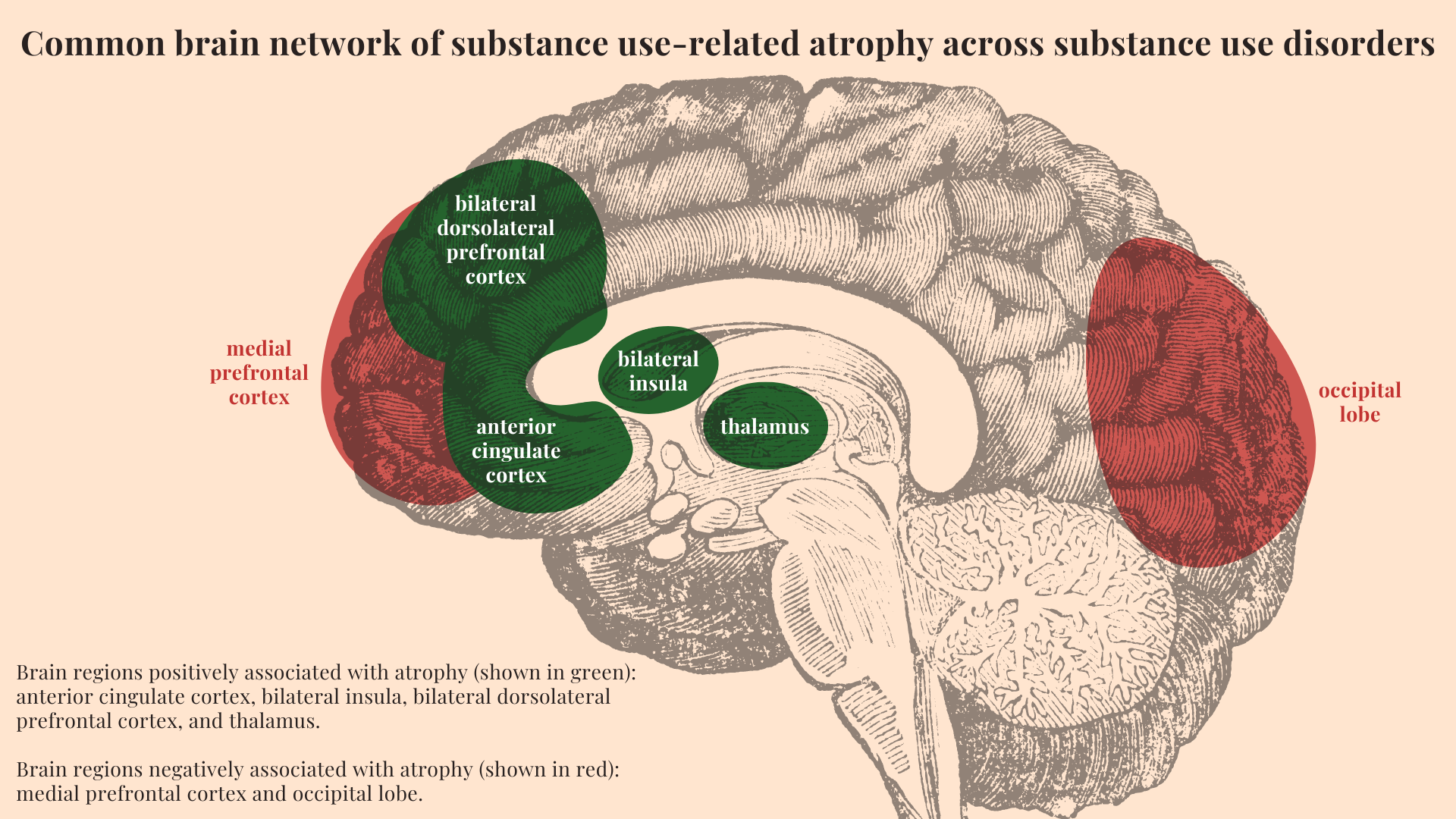
Figure. Common brain network of substance use-related atrophy across substance use disorders. Brain regions positively associated with atrophy (shown in green): anterior cingulate cortex, bilateral insula, bilateral dorsolateral prefrontal cortex, and thalamus. Brain regions negatively associated with atrophy (shown in red): medial prefrontal cortex and occipital lobe. Click image to enlarge.
Why do these findings matter?
Across substance use disorders, brain atrophy is localized to a common brain network. This finding supports the Syndrome Model of Addiction and suggests that treatment for one substance may be effective with other substances. For example, group cognitive behavioral therapy (CBT) can help reduce cocaine, opioid, and polysubstance use. It would be interesting to learn the extent to which this network is implicated in behavioral addictions, such as Gambling Disorder.
Every study has limitations. What are the limitations in this study?
Neuroimaging approaches varied across the 45 included studies. Differences in software packages, image preprocessing, statistical analysis, and study sample demographics may have biased the data.
For more information:
If you are worried that you or someone you know is experiencing addiction, the SAMHSA National Helpline is a free treatment and information service available 24/7. For more details about addiction, visit our Addiction Resources page.
— Caitlyn Matykiewicz, MPH
What do you think? Please use the comment link below to provide feedback on this article.