


Functional Magnetic Resonance Imaging (fMRI) studies have been critical to understanding the neurological mechanisms of addictive behaviors. Previous smoking research has identified brain regions involved in the processing of positive smoking-related cues that induce craving (e.g. a picture of a lit cigarette; Engelmann et al., 2012). Other imaging studies have examined smokers’ responses to negative smoking-related cues (e.g. picture of a chronic smoker’s diseased lungs), in efforts to understand how smokers think about cues that discourage smoking (Jasinska et al., 2012). These studies have been limited because of their narrow focus on predefined brain regions. Today, the ASHES reviews a study that uses a whole-brain analysis to examine how smokers process positive and negative smoking-related cues (Dinh-Williams, Mendrek, Bourque, & Potvin, 2014).
Methods
- Researchers recruited 30 chronic smokers through online advertisements and hospital referrals. Participants smoked an average of 19.1(SD=5.7) cigarettes a day for an average of 15.9(SD=9.6) years, were not attempting to quit, and did not have any contraindications for MRI.
- Participants completed questionnaires assessing demographic information and smoking history. During the scanning session, participants viewed 25 negative and 25 positive smoking-related images.1
- Researchers used multi-level models to test brain activation differences in response to positive and negative cues.
Results
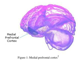
- There were multiple brain regions that were more responsive to positive as compared to negative smoking-related images, including the medial prefrontal area (see Figure 1) and the limbic system (see Figure 2).
- These brain regions are involved in mindful and comprehensive processing of stimuli (Lieberman, 2010), suggesting that the smokers did not process negative images as thoroughly as they did the positive images.
- Other brain regions were more responsive to negative smoking images than positive smoking images; however, these regions are not involved in mindful processing.

Figure 1. Medial prefrontal cortex. Click image to enlarge.

Figure 2. The Limbic System. Click image to enlarge.
Limitations
- The whole-brain analysis was exploratory and requires replication.
- Lack of a comparison group (e.g., non-smoking sample, treatment-seeking smokers, occasional smokers) makes it difficult to determine whether these findings are limited to specific groups of smokers.
Conclusion
Among chronic smokers, brain regions associated with thoughtfully thinking about a stimulus (i.e., limbic and medial prefrontal areas) were more responsive to viewing positive (vs. negative) smoking-related images. These results suggest that smokers are relatively less engaged in processing the negative effects of smoking. The researchers speculate that smokers might need to be taught to think more carefully about smoking’s negative effects in order to quit. Future research might further elucidate this phenomenon, including the voluntariness of this response and its role in continued smoking despite negative consequences.
— Aaron Lim
References
Dinh-Williams, L., Mendrek, A., Bourque, J., & Potvin, S. (2014). Where there’s smoke, there’s fire: The brain reactivity of chronic smokers when exposed to the negative value of smoking. Progress in Neuro-Pychopharmacology and Biological Psychiatry, 50, 66-73.
Engelmann, J.M., Versace, F., Robinson, J.D., Minnix, J.A., Lam, C.Y., Cui, Y., Brown, V.B., & Cinciripini, P.M. (2012). Neural substrates of smoking cue reactivity: A meta-analysis of fMRI studies. NeuroImage, 60, 252-262.
Jasinska, A.J., Chua, H.G., Ho, S.S., Polk, T.A., Rozek, L.S., & Stretcher, V.J. (2012). Amygdala response to smoking-cessation messages mediates the effects of serotonintransporter gene variation on quitting. NeuroImage, 60, 766-773.
Lieberman, M.D. (2010). Social cognitive neuroscience. In S.T. Fiske, D.T. Gilbert, G. Lindzey, (Eds.), Handbook of social psychology, Vol 1 (5th ed.) (pp. 143-193). Hoboken, NJ: John Wiley & Sons Inc.
What do you think? Please use the comment link below to provide feedback on this article.
1 Positive and negative images were randomized in blocks of 5. Each image was visible for 4 seconds, with a blank screen between each picture that lasted up to 1.5 seconds.
2 Image obtained from http://www.niaaa.nih.gov/research/niaaa-research-highlights/chronic-drinking-may-alter-brain-increase-ptsd-risk
3 Blausen.com staff. “Blausen gallery 2014”. Wikiversity Journal of Medicine.
doi:10.15347/wjm/2014.010