


Although pathological gambling falls under the category “impulse control disorder” in the Diagnostic and Statistical Manual of Mental Disorders – IV (American Psychiatric Association, 1994), there is some speculation that the brain structures involved in gambling are similar to those involved in addictive disorders, such as cocaine dependence (Bachs & Waal, 2003; Breiter, Aharon, Kahneman, Dale, & Shizgal, 2001). Recent innovations in neuroscience permit researchers to identify neural systems associated with disorders and to compare neurological profiles. Uncovering the neural systems associated with pathological gamblers has the potential to influence future treatment programs. This week the WAGER reviews Potenza et al. (2003) who examined the brain activity of pathological gamblers and controls while they watched three different video scenarios presenting gambling, happy or sad themes.
Through advertisements, the researchers recruited 13 male control participants and 14 male pathological gamblers. The pathological gamblers met the DSM-IV criteria for the disorder. All participants were free of any DSM-IV Axis I disorder except nicotine dependence. Although researchers could not exclude nicotine dependence, participant groups did not differ significantly in smoking status (1). Participants viewed three videotaped scenarios corresponding to three themes: happy, sad and gambling.
In the videos, actors talked directly to the camera, communicating either a happy scene (i.e., involving a wedding or an unexpected friend), a sad scene (i.e., involving a parental divorce or a relative’s death), or a gambling scene (i.e., involving a character who feels stressed, receives cash or a check, and invites the participant to gamble). The gambling scene included psychological cues for gambling (as specified by the author) such as problems at work or home, frustration, a period of unoccupied time, and the receiving of a check or a bonus. During the video, participants were instructed to press a button at the onset of a self-perceived emotion (e.g., happiness, sadness) or motivation (e.g., desire to eat, drink). Participants were not obliged to press the button.
This WAGER focuses on the data collected during baseline (i.e., before the video), and during the early part of the video (i.e., the period before participants pressed the button, or if they did not press the button, the first 45 seconds of the video). Researchers removed participants who pressed the button too quickly or those who experienced technical problems from the sample.
During this early part of the gambling scene, the viewer saw some of the following events depending on when or if s/he pressed the button: the actor “reports feeling criticized, acts stressed/annoyed regarding household chores, and criticizes others”; the actor has a period of free time and receives an unexpected sum of money (Potenza et al., 2003, p. 831). Researchers believed that this period would correspond to “early responses, before subjective awareness of internal state change” (Potenza et al., 2003, p. 830). MRI brain images were taken throughout this time period.
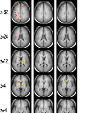
Figure 1. Highlighted in color are regions in which subjects with pathological gambling (n=10) differ at p<0.005 from control subjects (n=11) during viewing of the initial portions of gambling (A), sad (B) and happy (C) scenarios (E0-B1 comparisons). Red/yellow color highlights regions in which control subjects show more activation or less deactivation as compared with subjects with pathological gambling. Blue/purple color highlights regions in which control subjects show less activation or greater deactivation as compared with subjects with pathological gambling. Talaraich z levels are indicated. The left side of the brain is displayed on the right.

Images provided courtesy of Marc N. Potenza, MD, PhD, and the Yale University School of
Medicine.
In the gambling scenario, researchers found differences between pathological gamblers and controls when participants watched part of the video but had not begun to subjectively respond to the material, emotionally or motivationally (Figure 1). In this period, fMRI scans revealed decreased activity in the cingulate gyrus as well as in several other areas of the cortico-basal-ganglionicthalamic system in PG subjects compared to these same areas in control subjects. Subject groups expressed no significant differences in brain activity during the happy or sad scenarios.
A limitation of the study design includes the assumption that, before participants press the button, they are not subjectively responding to the visual information. Instead, participants might have been subjectively processing visual information but withholding the button press until they felt a sufficiently strong emotional or motivational response. Next, although researchers defined a variety of circumstantial cutes (e.g., reports feeling criticized, acts stressed/annoyed regarding household chores, etc.) as a gambling cues, these might not influence all pathological gambling participants similarly. Finally, like circumstances in other laboratory experiments, an individual’s response to happy, sad, and gambling videos might differ from an individual’s response to these situations in real life. For example, distractions like being placed in an MRI, noises that accompany image processing, and the absence of human interaction might affect a participant’s emotional and cognitive response.
These results, however, provide evidence supporting the view that pathological gamblers have a significantly different initial response to gambling-related scenarios than controls. Previous research exploring obsessivecompulsive disordered (OCD) participants (Saxena & Rauch, 2000) and cocaine dependent participants (Wexler et al., 2001) during the same period before subjective emotional/motivational response to disorder-relevant stimuli revealed an increase in the cortico-basal-ganglionicthalamic loop of the brain. The results of the present study show that pathological gambling participants exhibit a decrease in the same area. It is thought that this corticobasal-ganglionic-thalamic circuit perhaps signals a hyper activation of a response inhibition in OCD and cocaine dependent subjects as opposed to minimal response inhibition in pathological gambling subjects. This minimal response among pathological gamblers suggests that they might evidence similar neurological activity to non-drug disorders (e.g., trichotillomania, kleptomania ) that feature impaired impulse control (i.e., little response inhibition). These findings suggest that future treatments that specifically target the neurobiology of impulse control among pathological gamblers might reduce problematic gambling behavior.
Comments on this article can be addressed to Michael Stanton.
Notes
1 Researchers used the Fagerstrom Test for Nicotine Dependence (PG subjects: 2.10 ± 2.81; controls: 0.46 ± 1.51; x21, 19 = 2.88; P = .19)
References
American Psychiatric Association. (1994). DSM-IV: Diagnostic and statistical manual of mental disorders (Fourth ed.). Washington, D.C.: American Psychiatric Association.
Bachs, L., & Waal, H. (2003). [Naltrexone in the treatment of addiction]. Tidsskrift for Den Norske Laegeforening, 123(12), 1665-1667.
Breiter, H. C., Aharon, I., Kahneman, D., Dale, A., & Shizgal, P. (2001). Functional imaging of neural responses to expectancy and experience of monetary gains and losses. Neuron, 30(2), 619-639.
Potenza, M. N., Steinberg, M. A., Skudlarski, P., Fulbright, R. K., Lacadie, C. M., Wilber, M. K., et al. (2003). Gambling Urges in Pathological Gambling. Archives of General Psychiatry, 60(8), 828-836.
Saxena, S., & Rauch, S. L. (2000). Functional neuroimaging and the neuroanatomy of obsessive-compulsive disorder. Psychiatric Clinics of North America, 23(3), 563-586.
Wexler, B. E., Gottschalk, C. H., Fulbright, R. K., Prohovnik, I., Lacadie, C. M., Rounsaville, B. J., et al. (2001). Functional magnetic resonance imaging of cocaine craving. American Journal of Psychiatry, 158(1), 86-95.