


Neuroimaging is now a routine procedure, and "MRI" and "CAT-Scan" have become entrenched in the medical volcabulary of the lay person. But as these techniques become more common, the ways they are used in researching become more uncommon. Rogers, Owen, Middleton, Williams, Pickard, Sahakian, and Robbins (1999) used PET (positron emission tomography) and MRI (magnetic resonance imaging) technology to examine how participation in a gambling task affected patterns of blood flow in the brain. Their finding of localized "peak" regions of activity may shed light on the parts of the brain that are involved in the decision-making processes used by gamblers. 
Eight male subjects participated in the present study. None had a history of psychiatric illness or neurological disorders. The investigators developed a computerized gambling task that required subjects to bet points on finding a hidden yellow token. The task was designed so that subjects had to choose between small, likely rewards or larger, unlikely rewards. After being injected with a tracer compound, subjects were scanned as they worked on the task. [For more information about PET scans, click here]
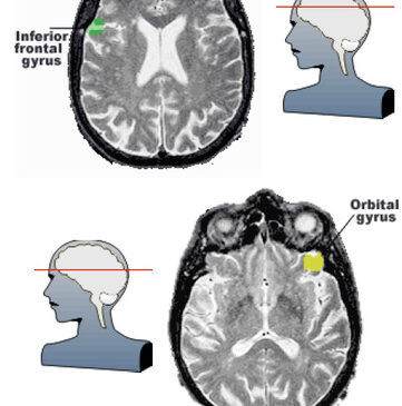
After the experiment, the investigators analyzed the PET and found that certain parts of the brain seemed to "light up" during the task. By looking at these areas, they were able to measure changes in regional cerebral blood flow (rCBF) and localize specific "peak" areas. They found changes in several brain regions, including the middle frontal gyrus, orbital gyrus, and the inferior frontal gyrus. The locations are indicated in the transverse MRI images at left. [Additional information on brain anatomy can be found in the Whole Brain Atlas, from which these images are taken]
It is true that the simulated gambling task used in this study may vary considerably from the experience of "real life" gambling. However, knowing which brain areas are involved in cognitive processes related to gambling may help future researchers better understand possible neuropathologies associated with compulsive
Source: Pasternak IV, A.V. & Fleming, M.F. (1999). Prevalence of gambling disorders in a primary care setting. Archives of Family Medicine, 8, 515-520.
The WAGER is funded, in part, by the National Center for Responsible Gaming, the Massachusetts Department of Public Health, the Andrews Foundation, the Addiction Technology Transfer Center of New England, the Substance Abuse and Mental Health Administration Services, and the Center for Substance Abuse Treatment.
Kevin Barnes December 7, 2011
Kevin Barnes
I really liked your blog article.Really looking forward to read more. Will read on…
Branden Feagin January 12, 2012
Branden Feagin
Very good blog article.Really thank you! Want more.